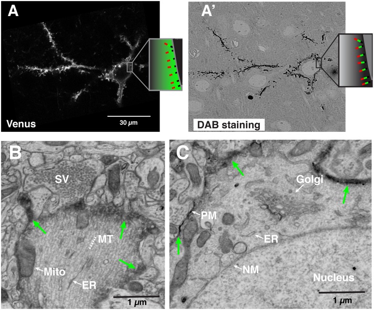
Figure 2.
Correlative LM-EM labeling of single neocortical neurons. (A,A’) Correlated images of Venus imaged by confocal microscopy (A) and DAB signal taken by the electron microscope (A’) from a single neuron labeled with APEX2-Venus-CAAX protein. (Inset; Schema illustrating the topology of the AVC protein on the plasma membrane; Red, Apex2. Green, Venus. Black, CAAX motif). The thickness of the section imaged in (A’) was 1 µm. (B–D) Single scanning electron micrographs showing the intracellular structures of a single labeled pyramidal neuron in layer II/III at P25. Green arrows indicate the plasma membrane labeled with APEX2-mediated DAB staining. (B) Primary apical dendrite. Arrowheads indicate microtubules (MT). (C) Cell body and its resident organelles: the Golgi apparatus, Nuclear membrane (NM), and the plasma membrane (PM) labeled with electron-dense DAB from APEX2. Abbreviations: SV, synaptic vesicle. MT, microtubule. Mito, mitochondria. ER, endoplasmic reticulum.

