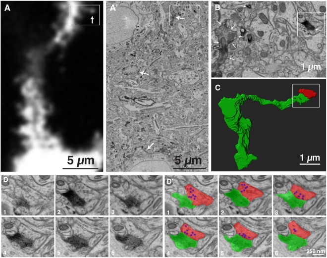
Figure 3.
Correlative LM-EM imaging of a dendritic spine and its presynaptic partner. (A) A single confocal light microscopy image of a dendrite labeled with AVC. The arrow indicates the spine reconstructed in (C,D). (A’) An electron micrograph corresponding to the area show in (A). The area surrounded by a rectangle is shown in (B). (B) Magnification of the area indicated by a rectangle in (A’). The area surrounded by a rectangle is shown in (D). Arrows in (A’,B) indicate AVC labeled neuron. (C) The structure of a spine shown in (A) reconstructed from serial electron micrograph. Green; labeled spine, Red; its presynaptic partner. (D) Serial electron micrograph of the area indicated in (B). (D’) The DAB staining is highlighted with green and the presynaptic partner of the labeled spine is highlighted with red. Synaptic vesicles in the presynaptic bouton are highlighted with blue.

