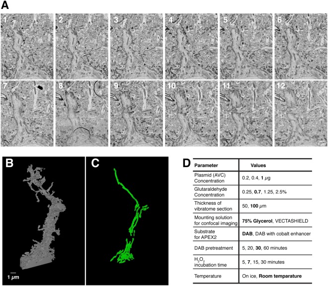
Figure 4.
3D-SEM reconstruction of the primary apical dendrite of single AVC-labeled pyramidal neuron and its subcellular features such as mitochondria. (A) Montages of serial electron micrographs including the DAB labeled pyramidal neuron shown in Fig. 1D,E. (see Supplemental Movie 3). (B,C) 3D reconstruction of the plasma membrane (B) and the mitochondria (C) of its labeled apical dendrite (see Supplemental Movies 4 and 5). To find out the region of interest, 256 serial sections were imaged with 30 nm/px resolution. The region of interest was re-imaged with 3 nm/px resolution through 103 serial sections and reconstructed. (D) Parameters tested to optimize the DAB staining and preservation of the membrane structures for SSEM. Bold indicates the optimal conditions used in the paper.

