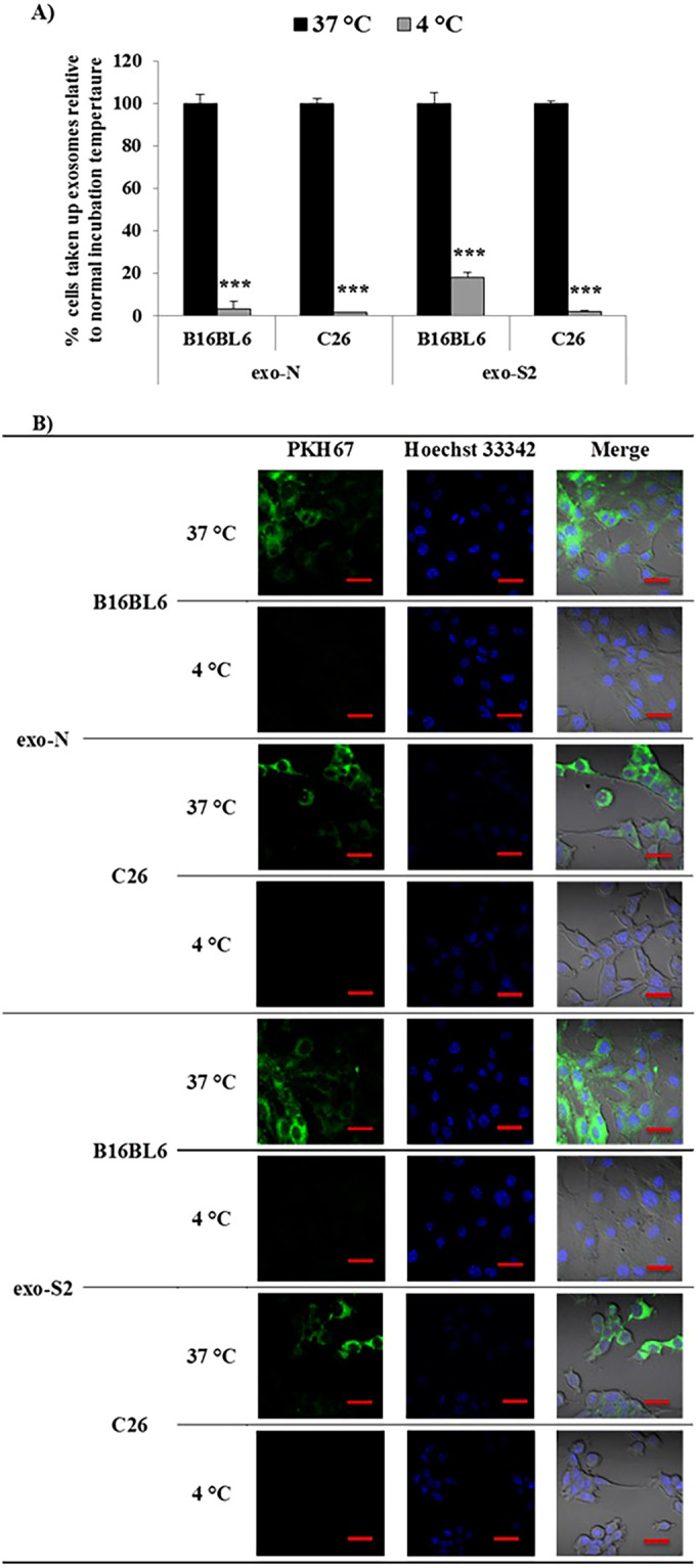
Figure 4.

Effect of temperature on exosome (EV) uptake by donor cells (B16BL6) and allogeneic cells (C26). Labeled samples of exo-N and exo-S2 were incubated with different cancer cell lines at 4 °C or 37 °C. After 2 h incubation, cancer cells were harvested for analysis by flow cytometry (A) or imaged by laser scanning confocal microscope after staining the DNA core with Hoechst 33342 (B). All data represent the mean ± SD (A) and one set (B) of triplicates. An unpaired t test was applied for each value relative to untreated cancer cells (***p < 0.001). Exosomes (EVs) were labeled with PHK67 (green) and the DNA core was stained with Hoechst 33342 (blue). Scale bar indicates 20 µm.
