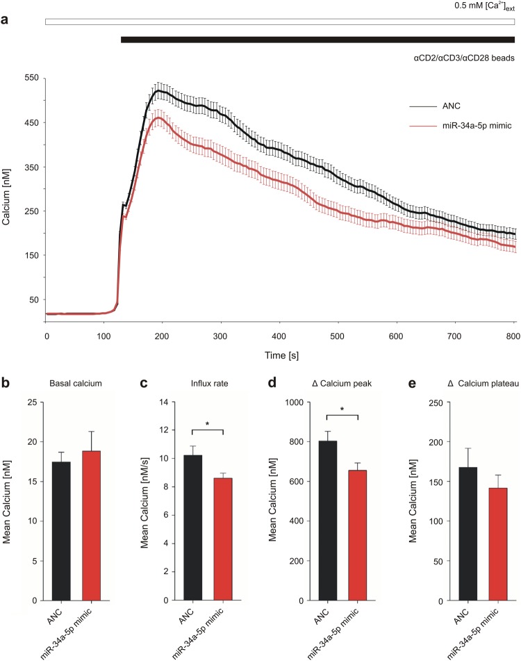
Fig. 6. Impact of miR-34a-5p overexpression on calcium signaling in Jurkat cell activation.
Jurkat cells were transfected for 48 h either with non-targeting control (ANC) or synthetic miR-34a-5p mimic. Respective Ca2+ imaging was performed in eight independent experiments on 3 days of measurement. Intracellular Ca2+ was sensed by Fura-2-AM fluorescent dye and SOCE was induced by αCD2/αCD3/αCD28 beads, while providing external Ca2+ solution. a Intracellular calcium concentration was determined over time of measurement and is shown as mean of all tested cells (ANC: n = 407; miR-34a-5p: n = 277). b–e Functional sections of the imaging procedure were evaluated as mean for each of the eight experiments respectively and are shown as mean for all experiments with corresponding SEM. Statistical evaluation was performed using Student’s t-test. A normal distribution of the data was assumed. (*p ≤ 0.05)

