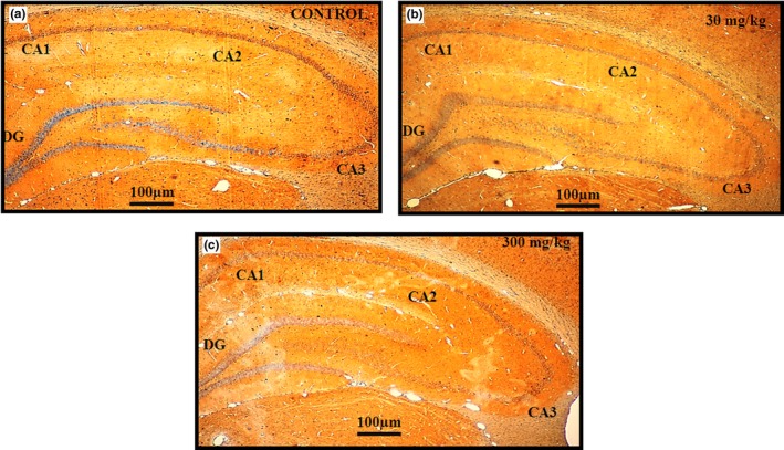
Figure 9.

HRP/DAB staining – Immunohistochemical analysis for qualitative detection of the expression of the GABAA α1 receptor subunit distribution in sections of rat hippocampus sub regions CA1, CA2, CA3, and DG in control (n = 5) (A), 30 mg/kg (n = 5) (B), and 300 mg/kg (n = 5) (C) groups of rats following 14 days of administration of the extract of Centella asiatica. Images (A, B, C) show moderate expression (++), with no significant difference between the control and treatment groups. The cells and tissues were labeled with the chromogen 3‐3‐diaminobenzidine (DAB). Three blinded investigators agreed on their observations. Scale bar = 100 μm
