
Figure 2.
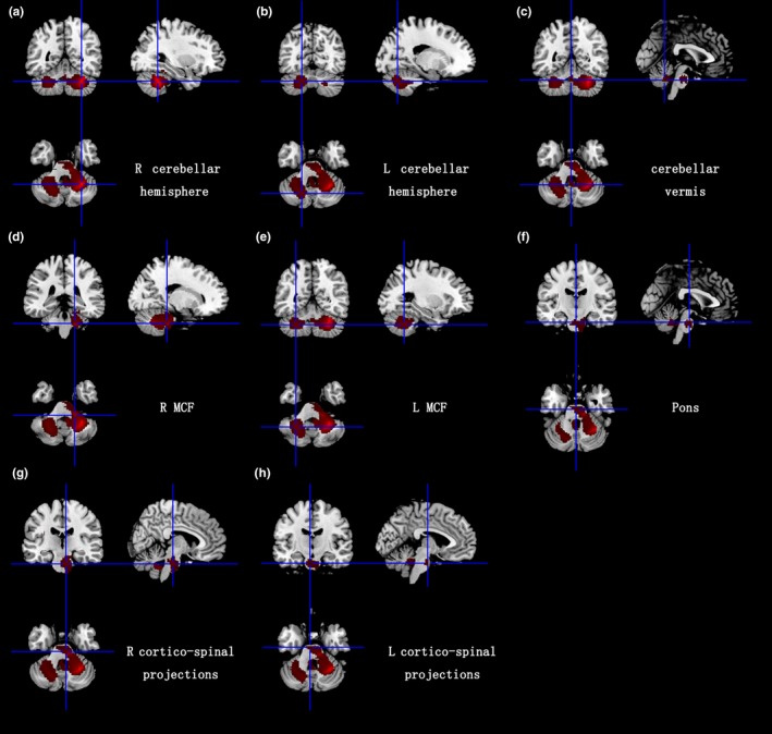
White matter atrophy in bilateral cerebellar hemispheres, cerebellar vermis, middle cerebellar peduncles, pons, and bilateral cortico‐spinal projections. R: right; L: left; MCF: middle cerebellar peduncles
Official websites use .gov
A
.gov website belongs to an official
government organization in the United States.
Secure .gov websites use HTTPS
A lock (
) or https:// means you've safely
connected to the .gov website. Share sensitive
information only on official, secure websites.
White matter atrophy in bilateral cerebellar hemispheres, cerebellar vermis, middle cerebellar peduncles, pons, and bilateral cortico‐spinal projections. R: right; L: left; MCF: middle cerebellar peduncles