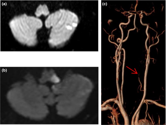
Figure 4.

DWI image of a 61‐year old man with transient isolated vertigo showed left cerebellar infarction (a), his symptoms deteriorated 20h after onset and repeat MRI showed medullary infarction (b), the left vertebral artery was narrow and segmental absent (red arrow, c)
