
Figure 2.
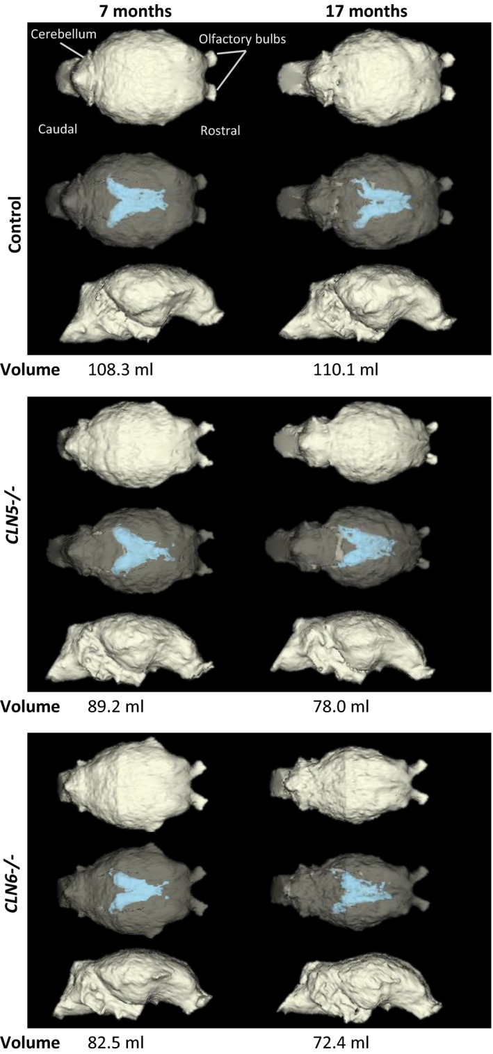
Representative 3D models of cranial volumes of a CLN5 and a CLN6 affected sheep and a control. Computer tomography (CT)‐based three‐dimensional (3D) modelling of intracranial volumes (ICV). In both CLN5 − / − and CLN6 − / −, the loss of ICV and enlargement of lateral cerebral ventricles (blue) were evident at 7 months of age and were pronounced at 17 months compared with the healthy control. The top row of each block shows the dorsal view, the middle row shows the semi‐opaque dorsal view so that the lateral ventricles are visible, and the bottom row shows the lateral view