Figure 1.

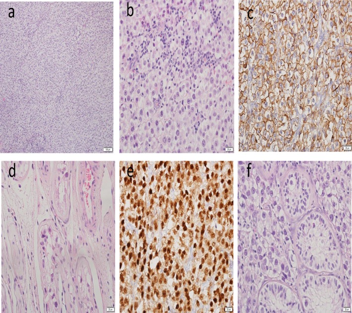
Cell morphology of seminoma by H&E staining and immuno‐stains. (a) General view × 10 (b) General view × 40 (c) c kit × 40 (d) ITGCN outside tumor × 40 (e) OKT4 × 40 (f) tumor with ITGCN at periphery × 40

Cell morphology of seminoma by H&E staining and immuno‐stains. (a) General view × 10 (b) General view × 40 (c) c kit × 40 (d) ITGCN outside tumor × 40 (e) OKT4 × 40 (f) tumor with ITGCN at periphery × 40