Figure 1.
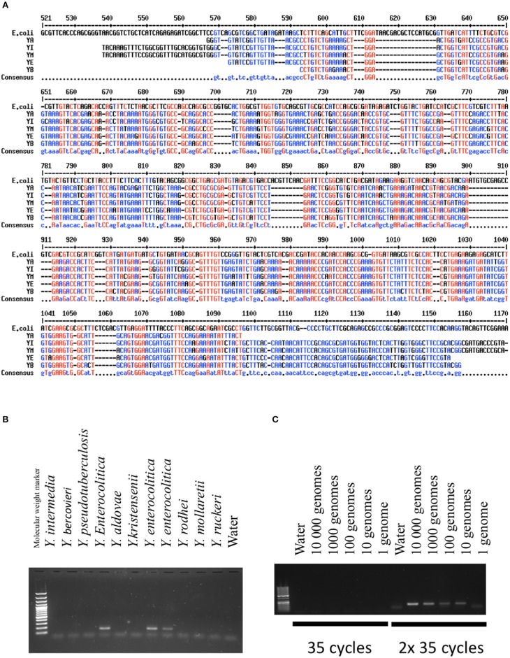
Validation of the PCR methods. (A) Comparison of GyrB sequences between different Yersinia species and E.coli. In red are indicated the nucleic acids present in all species. In blue is indicated the consensus sequence (Corpet, 1998). YA, Yersinia aldovae; YB, Yersinia bercovieri; YE, Yersinia enterocolitica; YI, Yersinia Intermedia; YM, Yersinia Mollaretii. (B) Specificity of the PCR methods developed for Y. frederiksenii (GyrB). DNAs from several species of Yersinia were amplified with the PCR technique and loaded on an agarose gel. A band indicates the presence of the specific PCR product specific to Y. frederiksenii. (C) Sensitivity of the PCR method developed for Y. enterocolitica (GyrB). The quantity of amplified DNA is expressed as genome equivalents. The method was highly sensitive when two cycles of 35 amplifications were performed.

