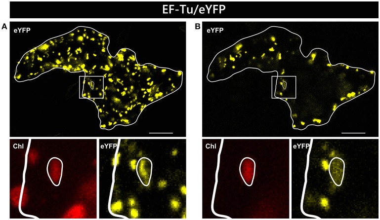
Figure 3.
Alternative visualization of EF-Tu/eYFP localization in epidermal cells. The same epidermal cell of Arabidopsis thaliana transiently transformed by particle bombardment with the EF-Tu/eYFP construct is shown either as maximum intensity projection of several single images representing the complete cell in z-axis (A) or as a single plane image from the same acquisition (B). In the areas below the overview pictures, separate images of eYFP and corresponding chlorophyll channels are shown at higher magnification. For further details see the legend of Figure 1.

