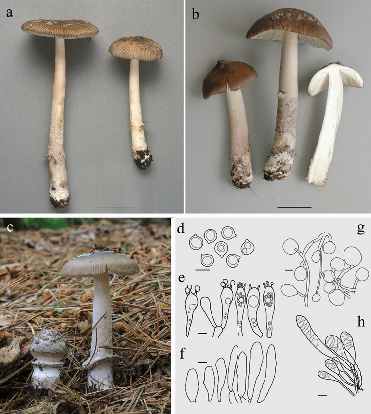
Figure 1.
Amanitarhacopus. a–c Basidiomes a CMMF002171(holotype), photograph by Yves Lamoureux b CMMF009640, photograph by Jacqueline Labrecque c HL016, photograph by Herman Lambert d–h Drawings of typical microscopic structures by Guy Fortin d Basidiospores e Basidia f Acrophysalides g Universal veil. h. Caulocystides. Scale bar: 3 cm (a, b), 10 µm (d, e), 20 µm (f–h).

