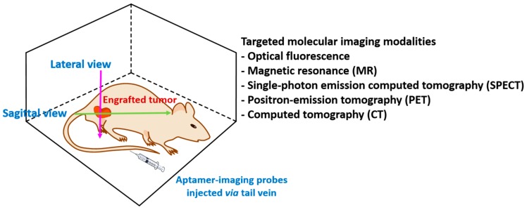
Figure 3.
Targeted in vivo imaging using aptamers. Mice possessing xenografted cancer cells are injected with aptamers labeled with imaging dyes. Non-invasive imaging modalities such as fluorescence, magnetic resonance (MR), single-photon emission computed tomography (SPECT), position-emission tomography (PET), or computed tomography (CT) are used for imaging on lateral or sagittal view.

