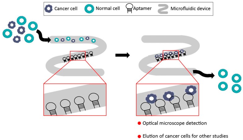
Figure 4.
Schematic illustration of a microfluidic device, showing a middle region with immobilized aptamers. After the injection of the sample, cancer cells are trapped by specific aptamer-cell interaction, whereas normal cells pass through the device. In this sense, cancer cells can be detected by optical microscope observation of the middle zone, and also eluted for future experiments. This device allows the isolation of cancer cells from a heterogeneous mixture of cells by selective cell-capture of immobilized aptamers.

