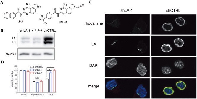
Figure 1.
Lamins were the efficacy targets of LBL1. (A) Chemical structures of LBL1 and LBL1-P. (B) LA expression was silenced by two independent shRNA constructs in DKO MEFs. (C) LBL1-P specifically labeled LA in DKO MEFs cells. The cells from part B were treated with LBL1-P and then subjected to the protocol of photo-cross-linking followed by click reaction with a rhodamine-N3. After click reaction, the cells were stained with anti-LA, and the cells were then analyzed by fluorescence microscopy. (D) DKO MEFs with silenced LA expression were resistant to LBL1. The cells from part B were treated with the indicated drug for 48 h. Then the viable cells were quantified by MTT assay. Data are presented as mean ± SEM (n = 5). * denotes P < 0.05.

