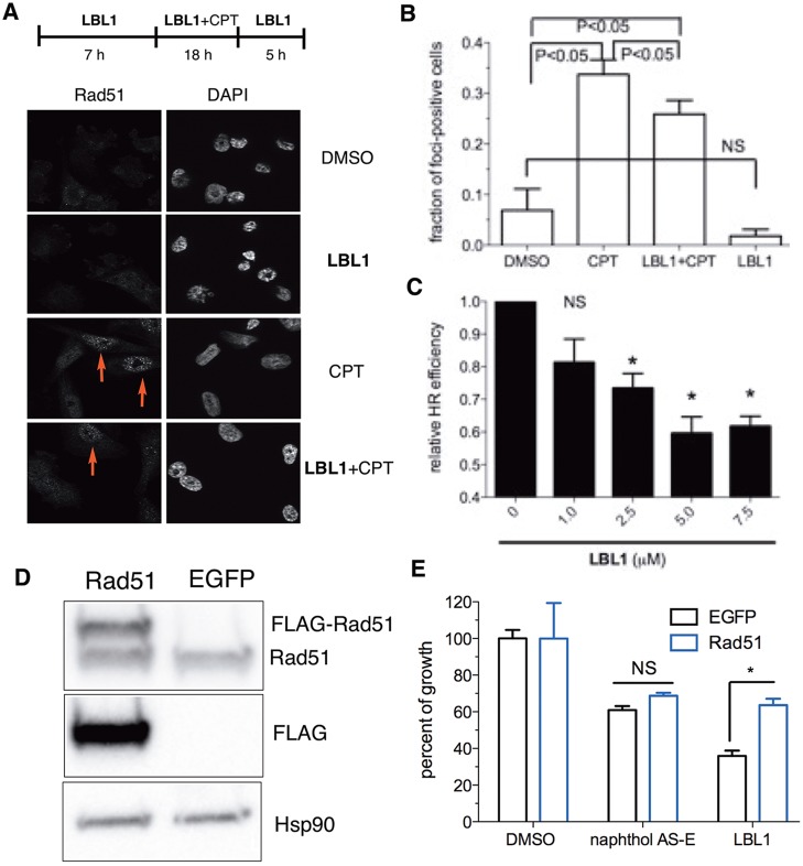
Figure 3.
LBL1 inhibited HR. (A) LBL1 inhibited Rad51 subnuclear foci formation stimulated by CPT. MDA-MB-231 cells were treated as described at the top. Then the cells were analyzed by immunofluorescence analysis. Representative images are shown. (B) Quantification of Rad51-foci-positive cells from part A. Data are presented as mean ± SD (n = 3, ∼100 cells were analyzed for each experimental condition). (C) LBL1 inhibited HR as assessed in a GFP-based reporter assay in MDA-MB-231 cells. The cells were transfected with a GFP-based HR reporter and DsRed as described in the Experimental Section. DSBs were induced by expressing I-SceI. Then the cells were treated with indicated concentrations of LBL1. The cells were then analyzed by flow cytometry. The ratio of GFP+/DsRed+ was registered as relative HR efficiency with vehicle-treated cells defined as 1.0 (n = 3). (D, E) Overexpression of Rad51 rescued LBL1’s antiproliferative activity in MDA-MB-231 cells. FLAG-tagged Rad51 was overexpressed in MDA-MB-231 cells, and the cell lysates were analyzed by Western blot with indicated antibodies. (E) The cells were treated with indicated drugs for 48 h. The cellular growth was quantified by the MTT assay. Data are presented as mean ± SEM (n = 3). * denotes P < 0.05.

