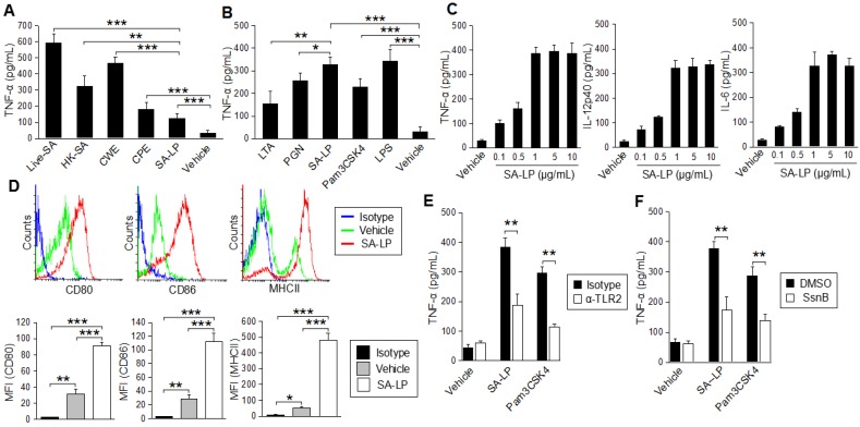
Figure 1.
SA-LP activates DCs in a TLR2-dominant manner. (A,B) BMDCs (1.0 × 10⁶) were stimulated with the S. aureus derived component or whole bacteria. Cultured medium was harvested after 24 hr of stimulation. The TNF-α production was measured by ELISA. (A) Live-SA (106), HK-SA (106), CWE, CPE, SA-LP (extracted from 106 of S. aureus). (B) LTA (10 μg/mL), PGN (10 μg/mL), SA-LP (10 μg/mL), Pam3CSK4 (500 ng/mL), LPS (100 ng/mL). (C) BMDCs (1.0 × 10⁶) were stimulated with SA-LP for 24 hr. Cultured medium was harvested, and the production of TNF-α, IL-12p40, and IL-6 were measured by ELISA. (D) Activation marker of SA-LP-stimulated BMDCs. BMDCs were stimulated by SA-LP (1 μg/mL) for 24 hr, then the stimulated cells were analyzed by flow cytometry to detect CD80, CD86, and MHCII. (E,F) BMDC stimulation under the blocking of TLR2 signaling. TLR2 was blocked with anti-mTLR2 mAd (10 μg/mL) (E), or inhibited with Sparstolonin B (100 μM) (F). The BMDCs were stimulated with SA-LP (1 μg/mL) for 24 h. Cultured medium was harvested and the TNF-α production was measured by ELISA. Live-SA; Live-S. aureus, HK-SA; Heat-Killed S. aureus, CPE; crude protein extract, CWE; cell wall extract, LTA; lipoteichoic acid, PGN; peptidoglycan, SA-LP; S. aureus crude lipoprotein. Data are shown as the mean and SD of at least three samples of a single experiment and are representative of at least three independent experiments. The Student’s t-test was used to analyze data for significant differences. Values of * p < 0.05, ** p < 0.01 and *** p < 0.001 were regarded as significant.

