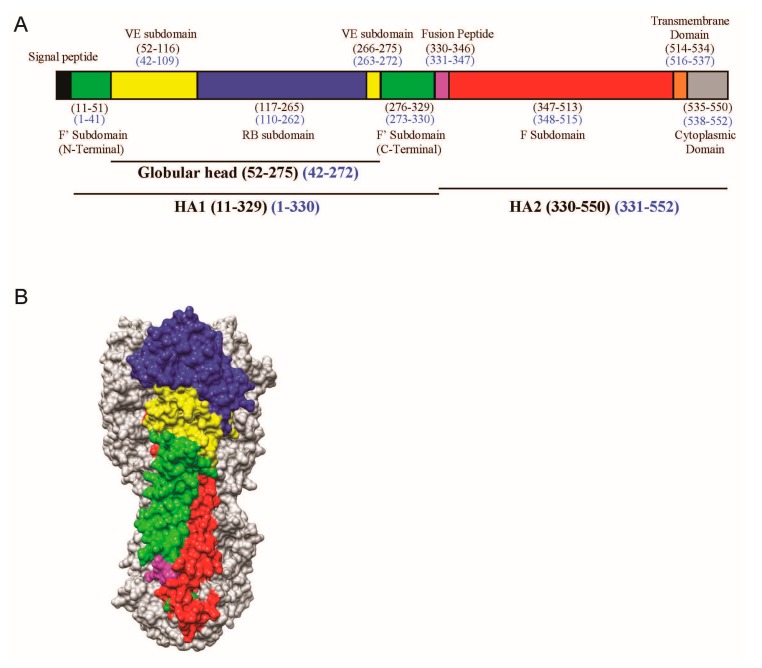
Figure 1.
(A) Schematic view of the H5 influenza viral haemagglutinin (HA) protein showing the different motifs present in HA. The stalk domain can be further divided into the F’ (green) or F (red) subdomains, whereas the globular head consists of the receptor binding (RB) subdomain (blue) and (vestigial esterase) VE subdomain (yellow), followed by the fusion peptide (magenta), transmembrane domain (orange), and cytoplasmic domain (grey). Numbering is indicated in the H3 (black) and H5 (blue) formats; (B) The corresponding segments in 3D are shown as a surface contour representation of one of the protomers of the H5 trimer from A/Vietnam/1203/04 (VN04) (PDB ID: 2FK0).

