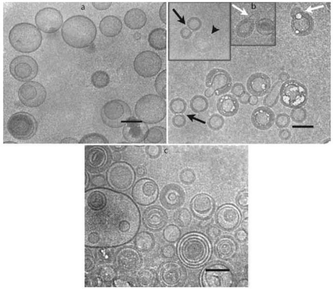
Figure 3.
Cryo-transmission electron microphotographs of 1,2-distearoyl-sn-glycero-3-phosphocholine (DSPC)/cholesterol (60:40, mol/mol) liposomes prepared in water (a) and after addition of 10% (w/v) lactose to the external solution (b). The microphotograph shown in (c) was collected after freezing the liposome dispersion shown in (b). The black arrows denote peanut shapes structures, white arrows collapsed outer membrane of double liposomes and arrowhead in the inset of (b) denotes a completely collapsed liposome. Bars 1⁄4 100 nm. Reproduced with permission from [31], Elsevier Inc., 2010.

