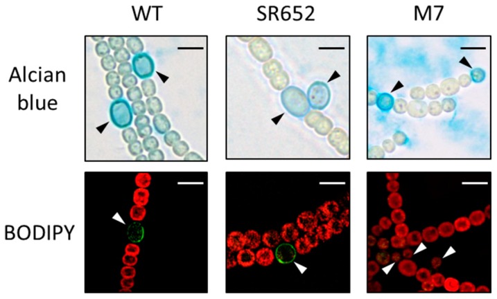
Figure 3.
Micrographs of the SR652 filaments in comparison to the WT and DevA-deficient mutant M7. Alcian blue stains exo-polysaccharides in blue; the green fluorescent dye BODIPY binds to neutral lipids. The red color indicates the cyanobacterial autofluorescence coming from the photosynthetic pigments. Heterocysts are indicated by triangles. Both staining procedures were performed after three days of nitrogen starvation. Bars, 5 μm.

