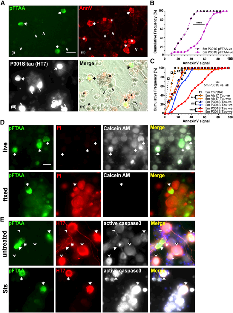
Figure 1. Living Neurons with pFTAA+Tau Filaments Aberrantly Expose PS by a Reversible ROS-Dependent Mechanism.

(A) Living DRG neurons from 5-month-old P301S mice with filamentous tau aggregates stained with (i) pFTAA (green) and (ii) AnnV-647 (red) (arrows) (asterisk denotes dead cell debris); (iii) same neurons fixed and stained for human tau (HT7 antibody). pFTAA-/HT7+ neurons do not stain with AnnV-647 (arrowheads). (iv) Nontransgenic (HT7-) neurons are AnnV-; images (i) and (ii) merged with phase contrast. Scale bar, 25 μm.
(B) Highersignal intensities ofAnnV-647 binding to pFTAA+ versus pFTAA- neurons in live cultures from 5-month-old P301S mice (****p < 0.0001). Cumulativefrequencyplot, 30 neurons perculture, n = 3 independent experiments. Kolmogorov- Smirnov test.
(C) Cumulative frequency plot comparing AnnV- 647 binding intensity values of HT7+ and HT7- neurons in the same cultures from 5-month-old P301S mice (***p < 0.001), 2-month-old P301S- mice (not significant [n.s.]), 5-month-old Alz17 mice (***p < 0.001), and HT7- 5-month-old wild- type C57 mice. AnnV staining of HT7+ neurons from 5-month-old P301S mice is significantly more intense than all others (***p < 0.001). Thirty neurons, n = 3 independent experiments. Kol- mogorov-Smirnov test with Bonferroni correction.
(D) pFTAA+ neurons from 5-month-old P301S mice are living cells. Top row: calcein-AM (white) and PI (red) staining showing exclusion of PI and retention of fluorescent calcein. Bottom row: after fixation, PI stains all cells, and there is no calcein retention. Data representative of n = 3 independent experiments. Scale bar, 30 mm.
(E) pFTAA+ neurons do not display PS, because of activation of caspase-3. Top row: pFTAA+ (green, arrows) and HT7+ (red)/pFTAA- neurons (arrowheads) showing basal intensities of active caspase-3 staining (white). Bottom row: staur- osporine (Sts) induces high levels of active cas- pase-3 in all the cells.
