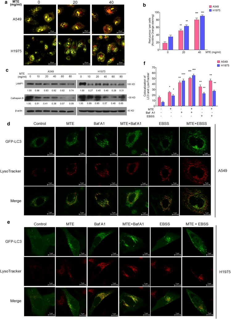
Fig. 4.
MTE suppressed autophagy by affecting lysosomal function. a Acidic vacuolar compartment in cells treated MTE with 0, 20 and 40 mg/ml (red puncta) were measured by acridine orange staining. b Numbers of red puncta in A549 and H1975 cells treated as in (a) were counted. Data represents mean ± SEM of at least 100 cells scored (*P < 0.05). c The protein level of LAMP1, Cathepsin B in treated cells were detected by Western blot, and protein expression levels were counted. Cells were treated with 0, 10, 20, 40, 60, 80 mg/ml MTE for 24 h. β-Actin was used as a loading control. d, e Colocalization of GFP-LC3 (green) and LysoTracker (red) were visualized on confocal microscope. GFP-LC3 stable A549 (d) and H1975 (e) cells were pretreated with 40 mg/ml MTE for 24 h following by Baf A1 (5 nM) or EBSS for another 4 h, and then incubated with LysoTracker for 90 min to be observed by confocal microscope. f Numbers of yellow (merge of green and red) puncta in cells treated as in (d, e) were counted. Data represents mean ± SEM of at least 100 cells scored. *P < 0.05, **P < 0.01, ***P < 0.005 vs control

