Fig. 3.
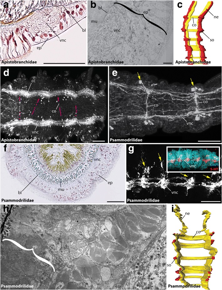
The VNC of Apistobranchidae and Psammodrilidae. Cross-sections (a, b, f, h), ventral (c-e, i) and lateral view (g) of the ventral cord in Apistobranchus tullbergi (a-d), Psammodrilus balanoglossoides (f, h, i) and Psammodrilus curinigallettii (e, g). Anterior is up (c, i) and left (d, e, g). Azan staining (a, f), TEM (b, h), anti-FMRFamide (d), anti-5-HT (e, g), and DAPI (g, inset) staining, and 3D-reconstruction (c, i). (a) The (vnc) in Apistobranchidae is intraepidermal. Giant fibers are absent. (b) Ultrastructural data verify a position of the (vnc) within the epidermis (ep). A basal lamina (bl) is present. (c) Apistobranchidae show a medullary arrangement of somata (so) and neuropil (ne). Somata-free areas are absent. Commissures (co) show a serial arrangement. (d) FMRFamide-immunoreactivity reveals the presence of two neurite bundles within the (vnc). Faint commissures (co) are visible. The location of the commissures (co) is marked with purple dots. (e) Anti-5-HT staining of the (vnc) in Psammodrilidae reveals presence of clustered somata (yellow arrowheads) and segmentally arranged commissures (co). (g) The (vnc) is intraepidermal. (g) Lateral view of the (vnc) verifies presence of immunoreactive somata clusters (yellow arrowheads). DAPI staining supports the anti-5-HT staining. (h) Ultrastructural investigations support the intraepidermal localization of the (vnc). The musculature (mu) is situated in subepidermal position. (i) A 3-D reconstruction in Psammodrilidae reveals the presence of clustered somata (so) along the (vnc). Segmentally arranged commissures (co) are present. bl, basal lamina; cc, circumesophageal connective; co, commissure; ep, epidermis; mu, muscle; ne, neuropil; pa, parapodia; so, somata; vnc, ventral nerve cord. Scale bars = 50 μm (a, e-g), 10 μm (b), 100 μm (d) and 2.5 μm (h)

