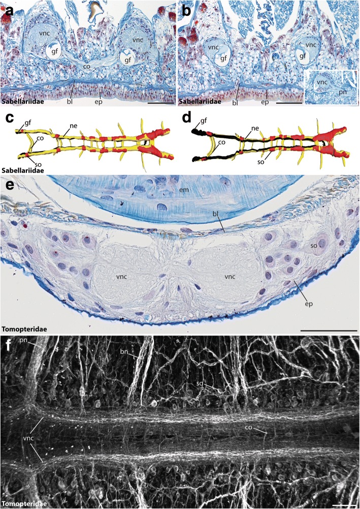
Fig. 5.
The VNC of Sabellariidae and Tomopteridae. Cross-sections (a, b, e) and dorsal (c) and ventral view (d, f) of the ventral cord in Sabellaria alveolata (a-d) and Tomopteris helgolandica (e, f). Anterior is right (c, d, f). Azan staining (a, b, e), 3D-reconstruction (c, d) and anti-α-tubulin staining (f). (a, b) The (vnc) in Sabellaria is situated in subepidermal position. The ventrally located giant fibers (gf) are visible throughout the entire length of the animal and numerous segmentally arranged commissures (co) are present. The inset shows an outgoing parapodial nerve (pn) branching of from the ventral nerve cord. (c, d) 3Dreconstructions reveal the presence of serial somata (so), bearing ganglia and commissures (co). (e) The (vnc) in Tomopteris is situated within the epidermis (ep) and consists of two distinct neurite bundles. (f) Immunohistochemistry reveals presence a medullary-like arrangement of somata (so), segmentally arranged commissures (co) and branching nerves (bn). Somata-free connectives are absent. bl, basal lamina; bn, branching nerve; co, commissure; em, esophageal musculature; ep, epidermis; gf, giant fiber; ne, neuropil; pn, parapodial nerve; so, somata; vnc, ventral nerve cord. Scale bars = 200 μm (a, b) and 100 μm (e, f)

