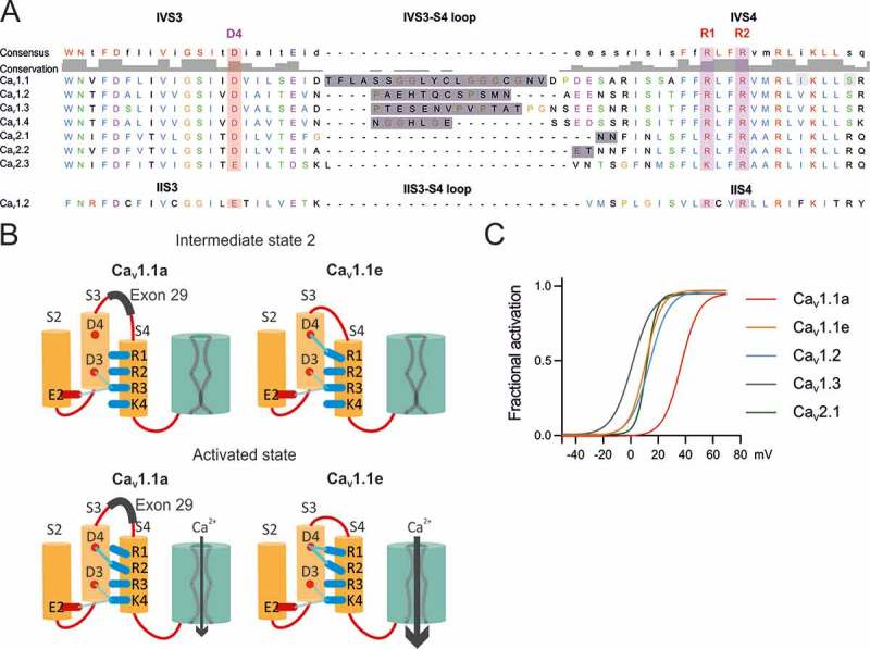
Figure 2.

Charge-neutralization of D4 in VSD IV of CaV1.3 affects voltage sensitivity and peak current density. (A) Domain structure of CaV1.3 indicating the location of D4 in the outer S3 helix of VSD IV. (B) GFP-CaV1.3 and mutant GFP-CaV1.3-ΔE32-D4N are targeted to triads of dysgenic (CaV1.1−/-) myotubes. Scale bar, 10 μm. (C) Representative calcium currents of GFP-CaV1.3-ΔE32 (blue) and mutant GFP-CaV1.3-ΔE32-D4N (orange) at 0 mV and + 20 mV. (D) I/V curves and (F) the scatter plot of the current density (I peak) show that D4N has a reduced current density. (E) Factional activation curves and (G) scatter plot of the V½ indicate that voltage sensitivity was reduced significantly by the charge neutralization of D4. (Mean± SEM; *p ≤ 0.05; **p ≤ 0.01; n = 9,10; Unpaired t-test).
