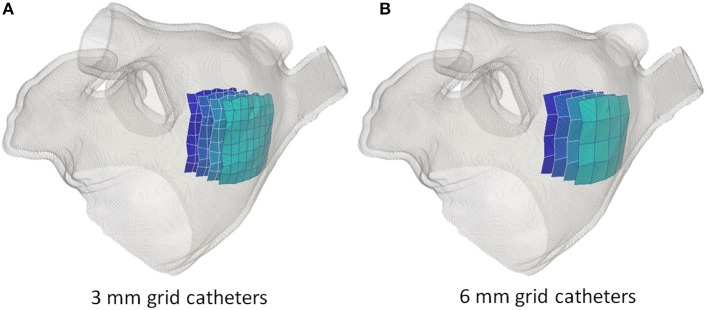
Figure 3.
Simulated grid catheters. In (A) the catheter with a 3 mm inter-electrode spacing. In (B) the catheter with a 6 mm inter-electrode spacing. In both cases, four catheters are illustrated corresponding to a distance to the atrial wall of 0 mm (lightest blue), 5, 10, and 15 mm (darkest blue).

