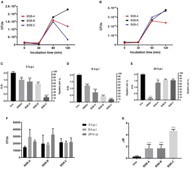
Figure 1.
Biological characterization of the three hypermucousviscous Klebsiella pneumoniae isolates (BSB-A; BSB-B, BSB-C). (A) Bacterial blood survival evaluation of CFUs (colony forming units) recovery after isolate incubation in blood for 30, 60, and 120 min. (B) Bacterial human serum survival evaluation of CFUs recovery after isolate incubation in serum for 30, 60, and 120 min. (C–E) Hep-2 cells viability after different periods of infection by K. pneumoniae isolates. (F) Bacterial invasion of Hep-2 cells by K. pneumoniae isolates evaluated by CFUs recovery after 3, 6, and 24 h of infection. (G) Nitric Oxide quantification in Hep-2 cells after 24 h of infection by K. pneumonia isolates. Uns: Unstimulated cells (not submitted to bacterial infection). Control: unstimulated cells (not submitted to bacterial infection). DMSO: positive control for citotoxicity. *p < 0.05; **p < 0.01; ***p < 0.001. All infected cells were compared to unstimulated cells (non infected cells).

