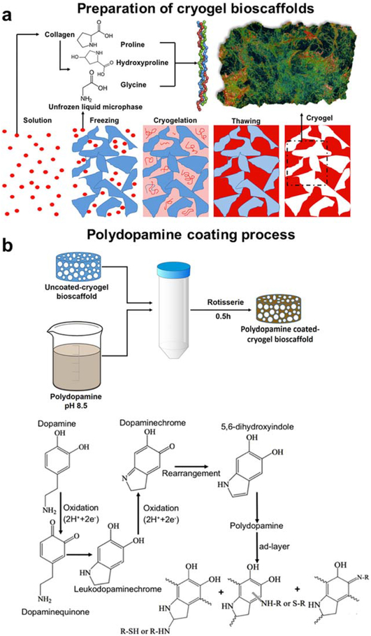
FIGURE 1.
Schematic illustration of the preparation of uncoated- and polydopamine coated-cryogel bioscaffolds: (a) Collagen was swollen overnight in HCl at 4°C and then the collagen dispersion was homogenized and centrifuged. After transferring the collagen slurry to a mold, NHS/EDC was added (depicted as the solution); The molds were kept in a freezer at −20°C for 24 h (depicted as the freezing) to complete the crosslinking process (depicted as the cryogelation). Next, bioscaf-folds were thawed at room temperature (depicted as the thawing); (b) Polydopamine was then coated by immersion of bioscaffolds into a dopamine solution (2 mg/ml in 10 mM Tris, pH 8.5) on a tube rotisserie; Schematic illustration of the polydopamine formation mechanism which involves the oxidation of catechol in dopamine to quinone by alkaline pH-induced oxidation.

