
Figure 5.
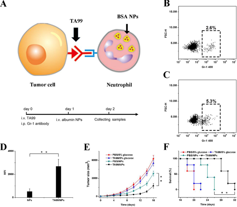
Neutrophils deliver NPs to tumors induced by antibody dependent cell-mediated cytotoxicity (ADCC). (A) Concept of NPs delivered by neutrophils to tumors mediated by ADCC. Percentage of neutrophils in tumor tissue with i.v. administration of (B) PBS and (C) TA99 at 48 h. The samples were single cell suspensions prepared from mouse tumors 48 h after treatment with PBS or TA99 in PBS. The cell suspension was analyzed by flow cytometry. (D) MFI of NPs in neutrophils in tumor by flow cytometry 48 h after the injection of Cy5-BSA NPs or both TA99 (i.v. injected at 24 h) and Cy5-BSA NPs (i.v. injected at 48 h) (n = 3). Neutrophils were stained with Alexa-488 anti-mouse Gr-1 antibody, and nucleus was stained by DAPI. (E) Tumor size and (F) survival rates of mice bearing melanoma illuminated with a 660-nm laser 48 h after i.v. injections of vehicles, TA99, Ppa-loaded NPs, or both TA99 (i.v. injected at 24 h) and Ppa-loaded NPs. Copyright 2016, Wiley.[14]