Figure 2.
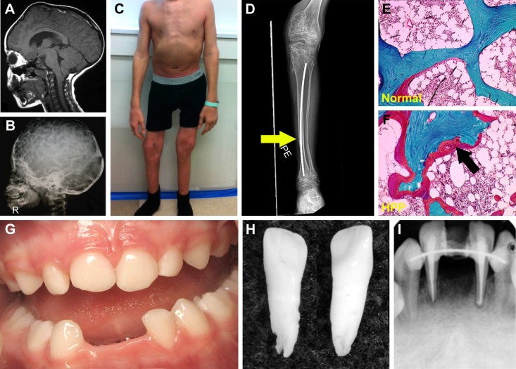
Skeletal and dental defects associated with hypophosphatasia.
Notes: (A) MRI of the skull of a 6-year-old individual with hypophosphatasia (HPP) exhibiting craniosynostosis and the resulting bregmatic bump. (B) Radiograph of a 4-year-old individual with HPP reveals hypomineralization of the cranial vault as seen by the “copper beaten” appearance of the skull. (C) Deformities of lower extremities with joint widening at knees and elbows in a 15-year-old boy with severe childhood HPP. (D) Knee radiograph of the same individual reveals hypomineralized bone, coarsened trabeculae, and an intramedullary rod in the tibia. A fracture line is seen at the diaphysis of the tibia (yellow arrow). (E) Goldner trichrome stain of normal iliac crest biopsy compared to (F) the same from an individual with adult HPP, showing accumulation of excessive osteoid (red layer indicated by black arrow) on the surface of the mineralized bone (green). (G) Oral photograph of a 2.5-year-old child with HPP exhibiting premature loss of primary lower incisors. (H) Primary incisors that spontaneously exfoliated from a child with HPP. Note the intact roots, a hallmark of HPP. (I) Oral radiograph of a 20-year-old individual diagnosed with odonto-HPP showing loss of secondary incisor, endodontic treatment after fracture, and splinting to try and stabilize remaining anterior teeth. Images in A and B are reprinted by permission from Springer Customer Service Centre GmbH: Springer Nature [Child’s Nervous System]; Neurosurgical aspects of childhood hypophosphatasia, Collmann H, Mornet E, Gattenlöhner S, Beck C, Girschick H. Copyright 2009.31 Images in E and F are reprinted from Bone, Vol /edition 54(1), Berkseth KE, Tebben PJ, Drake MT, Hefferan TE, Jewison DE, Wermers RA, Clinical spectrum of hypophosphatasia diagnosed in adults, Pages 21–27, Copyright (2013), with permission from Elsevier.36 Image in G is reproduced from Reibel A, Manière MC, Clauss F, et al. Orodental phenotype and genotype findings in all subtypes of hypophosphatasia. Orphanet J Rare Dis. 2009;4(1):6. Image in H is reproduced with permission from © 2017 American Society for Bone and Mineral Research. Whyte MP. Hypophosphatasia: enzyme replacement therapy brings new opportunities and new challenges. J Bone Miner Res. 2017;32(4):667–675.112 Image in I is reproduced with permission from Copyright © 2012, John Wiley and Sons. Rodrigues TR, Georgetti AP, Martins L, Pereira Neto JS, Foster BL, Nociti Jr FH. Clinical correlate: case study of identical twins with cementum and periodontal defects resulting from odontohypophosphatasia. In: McCauley LK, Somerman MJ, eds. Mineralized Tissues in Oral and Craniofacial Science: Biological Principles and Clinical Correlates. 1st ed. Ames, IA: Wiley-Blackwell; 2012.38

