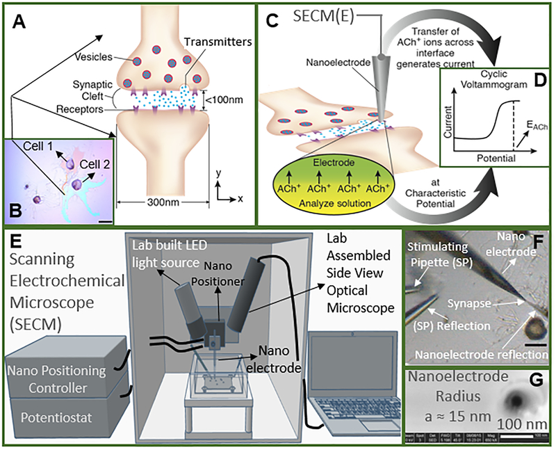
Figure 1.
Study of cholinergic neurotransmission at single synaptic cleft with nanoelectrode and scanning electrochemical microscope. (A) Illustration of synaptic cleft dimensions22–, 23, 24, 25, 26, 27. (B) Cultured living Aplysia pedal ganglion neurons used for the experiment, where the axon from cell 1 (pink) formed a synaptic connection with the body of cell 2. Scale bar: 200 μm. (C) A nanoITIES pipet electrode was positioned around the synaptic cleft to measure the concentration and release dynamics of acetylcholine (ACh+) simultaneously using amperometry; the positioning of the nanoelectrode was achieved using the scanning electrochemical microscope (Fig. E) with a spatial resolution of 5 nm. The zoom shows the nanoITIES formed at the tip of the nanoITIES pipet electrode, and ionic transmitter (ACh+) transfers across the interface, generating a current and thus getting detected. (D) Cyclic voltammogram corresponding to ACh detection, where the detection potential follows Nernstian equation, and a steady state transfer potential, EACh= - 0.48 V vs E1/2, TBA, selective for cholinergic neurotransmitter detection was used in amperometry to study its synaptic concentration dynamics (results shown in Figs. 2 and 3). (E) A Scanning Electrochemical Microscope (SECM) and a lab-built side view optical microscope were used for the positioning of the nanoelectrode around synapses with nm spatial resolution. The lab-built side view optical microscope provided rough positioning before the fine positioning of 5 nm spatial resolution with SECM. After SECM positioning (Supporting Information, figs. S9, S10), the optical microscopic view of the nanoelectrode and the synapse are shown in Fig. F, where it can be seen that it is very hard to locate the synapse by visual observation alone. The combined use of the side view optical microscope and nano-positioning platform, SECM, is critical. (F) A stimulating pipet was used to provide high concentration K+ stimulation. Reflection was used for the rough positioning of the nanoelectrode and stimulating pipet in the x, y and z axes by optical microscope, which was followed by the nanometer positioning of the nanoelectrode around the synapse achieved using nano-resolution SECM with details described in the supporting information. Scale bar: 150 μm. (G) High resolution scanning electron microscope (SEM) picture of the nanopipet tip with radius (a) to be around 15 nm.

