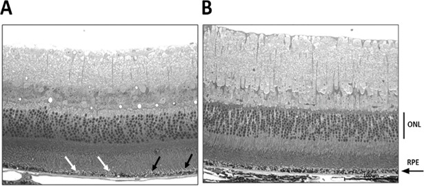
Fig 6. Light micrograph analysis.
Preliminary data showed increased melanosomes in the RPE layer in the zeaxanthin treated eyes (n = 2) (B) compared to untreated eyes of same age (n = 2) (A). Both images are from the same location in the posterior retina of each eye. (scale bar 100μm). In A, black the arrow indicates a region in which outer segments of photoreceptors appear detached from the RPE and the white arrow head indicates a region of hypopigmentation in the RPE.

