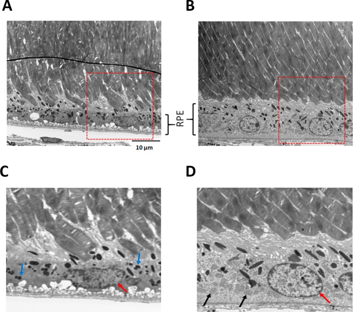
Fig 7. Improved ultrastructure of RPE.
Ultrastructure analysis showed preservation of photoreceptors and RPE in the zeaxanthin treated eyes (n = 2) (B and D), whereas the eye from untreated mice (n = 2) (A and C) revealed broken tips of photoreceptors outer segments. Mitochondria were visible in the treated RPE (black arrows in D) but were sparse in the untreated eyes. The accumulation of lipofuscin granules (small black dots indicated by blue arrows in C) were reduced in treated eyes. RPE of the untreated eye had visible nuclear alterations such as nuclear pyknosis whereas the RPE cells in treated eyes had a normal, round nucleus (red arrow).

