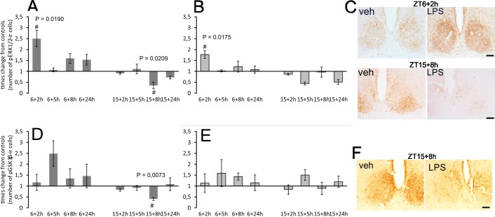
Fig 5.
Effect of acute systemic LPS administration on ERK1/2 (A, B) and GSK3β (D, E) phosphorylation within rat SCN. Adult rats were injected with LPS (1 mg/kg) either during the day at ZT6 or at night at ZT15 and sampled 2 h, 5 h, 8 h and 24 h later. Levels of immunopositive cells were assessed separately for the ventrolateral (A, D) and dorsomedial (B, E) SCN. Each column represents the mean of four values ± SEM. # P: Values of multiple t-tests with the Sidak-Bonferroni post-hoc test. The representative photomicrographs of coronal sections of the SCN demonstrate the intensity and distribution of pERK1/2 (C) and p GSK3β (F) in the control and LPS-treated animals when the control/LPS pairs showed statistically significant differences. Scale bar = 200 μm.

