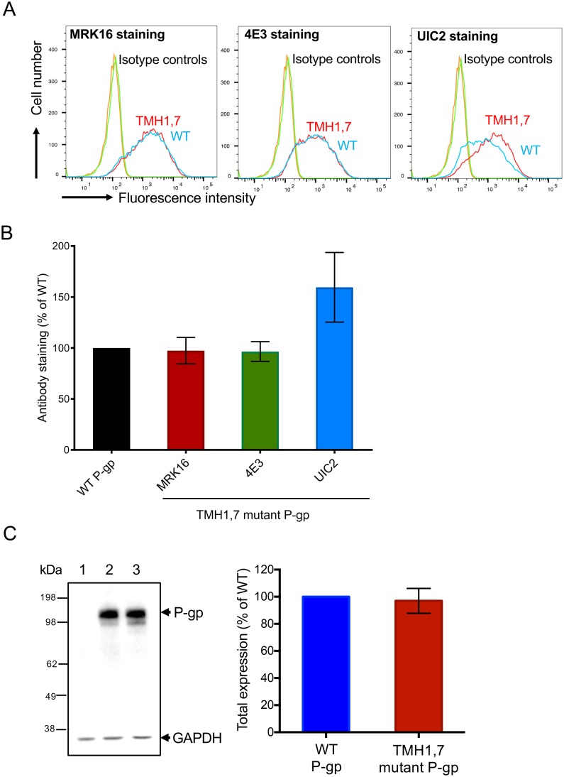
Fig 2. Total and cell surface expression of TMH1,7 mutant P-gp in HeLa cells.
HeLa cell surface expression of TMH1,7 mutant P-gp was determined by staining with monoclonal antibodies against human P-gp: MRK16, 4E3 and UIC2. (A) In flow cytometry histograms, the curves show the expression of TMH1,7 (red) and WT P-gp (blue). IgG2aκ isotype controls are also shown. (B) Using surface expression data from flow-cytometric analysis in (A) and expression of WT P-gp taken as 100%, the relative expression of TMH1,7 mutant P-gp was calculated. Five to eight independent replicates were quantified, and error bars show SD. (C) Western blot of lysates of HeLa cells transduced with BacMam baculovirus showing the total expression of WT and TMH1,7 mutant P-gp using C219 antibody. The lysate of 60,000 untransduced cells (lane 1), cells expressing WT-P-gp (lane 2) or TMH1,7 mutant P-gp (lane 3) was loaded. The GAPDH expression was used as a loading control. The experiment was repeated three times with independent transductions and relative expression of TMH1,7 mutant P-gp was calculated using WT level as 100%, shown in bar graph (right). Error bar shows SD of three experiments.

