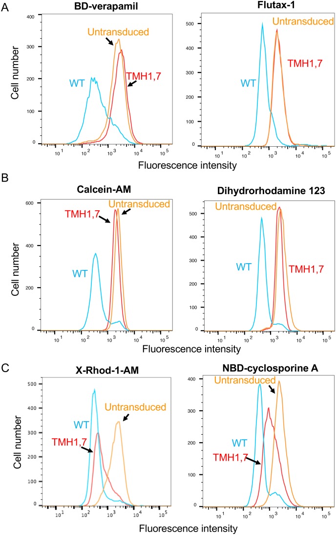
Fig 3. Characterization of the transport function of TMH1,7 mutant P-gp.
WT and TMH1,7 mutant P-gp were expressed on HeLa cells by the BacMam baculovirus-based transduction system. The transport of fluorescent substrates was analyzed using flow cytometry. Histograms indicate the transport of representative substrates that are not transported by TMH1,7 mutant P-gp, (A) BODIPY-verapamil (0.5 μM) and flutax-1 (5 μM); substrates that are partially transported (10–30% compared to WT) by TMH1,7 mutant P-gp, (B) calcein-AM (0.5 μM) and dihydrorhodamine 123 (1.3 μM); and substrates that are efficiently transported by TMH1,7 mutant P-gp, (C) X-Rhod-1-AM (0.5 μM) and NBD-cyclosporine A (0.5 μM). Fluorescence intensity of WT P-gp is shown in blue, TMH1,7 mutant P-gp as red and untransduced cells are orange traces in all histograms.

