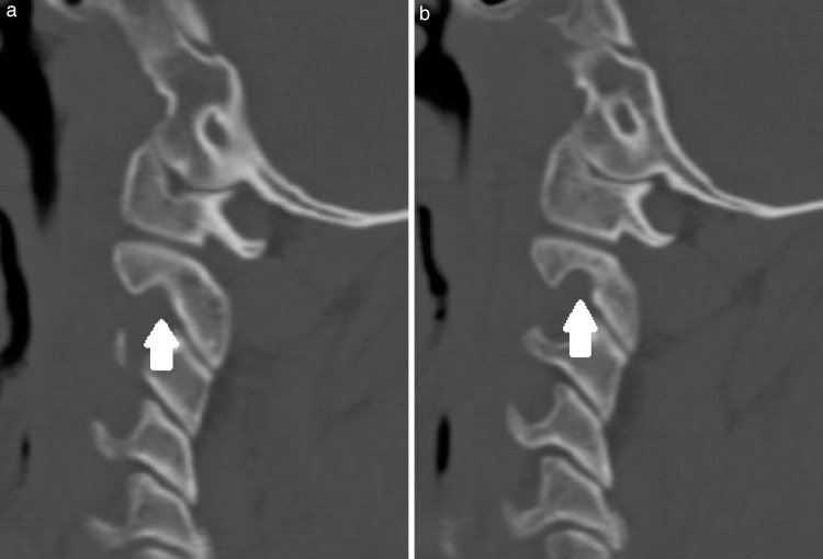
Figure 3.
(a) Identification of the vertebral artery foramen slice. A small portion of the superomedial aspect of the vertebral artery foramen can be seen in the C2 pedicle. (b) The next cut is 2 mm lateral and demonstrates a vertebral artery foramen with at least a full semicircular opening. This cut is deemed the vertebral artery foramen slice.

