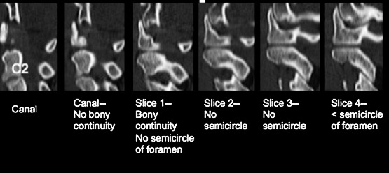
Figure 5.

Series of sagittal computed tomography cuts from the spinal canal through the pedicle. This patient has 4 slices of bony continuity that do not show the vertebral artery foramen. Thus, slices 1 through 4 represent the safe zone where a long pedicle screw can be placed.
