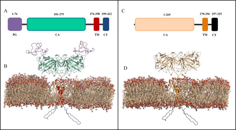
Fig. (2).
Schematic and structural models of CA IX* and XII. (A) CA IX domains include the proteoglycan-like domain (PG, purple), catalytic domain (CA, cyan), a transmembrane anchor (TM, red), and intracellular C-terminal domain (CT, blue). (C) CA XII domains include the catalytic domain (CA, wheat), the transmembrane domain (TM, gold), and intracellular C-terminal domain (CT, black). (B) Homodimeric membrane bound form of CA IX includes the extracellular PG and CA domains (purple and green), the TM anchor (red), and the CT domain (blue). (D) Homodimeric membrane bound form of CA XII includes the CA domain (wheat), the TM anchor (gold), and the CT domain (black). The TM domains are predicted to form helix-helix interactions within the lipid bilayer. The CT domains are predicted to exist as unstructured loops. * Structural model of CA IX provided by Brian Mahon [21].

