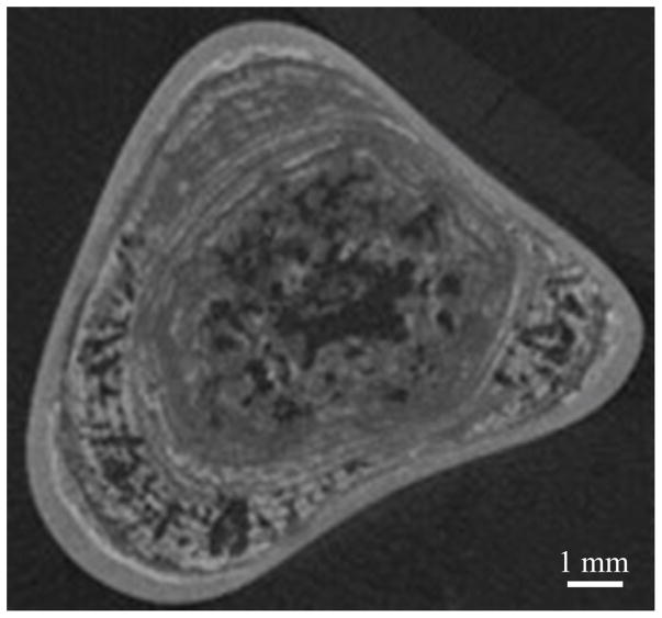
Figure 2.
Center microcomputed tomography (μCT) slice of a calcium oxalate monohydrate (COM) kidney stone. Gray structures in the image show the inorganic crystals; dark or radiolucent regions are indicative of the organic protein matrix and may contain pockets or trapped gas or microbubbles. Image courtesy of James C. Williams, Jr.

