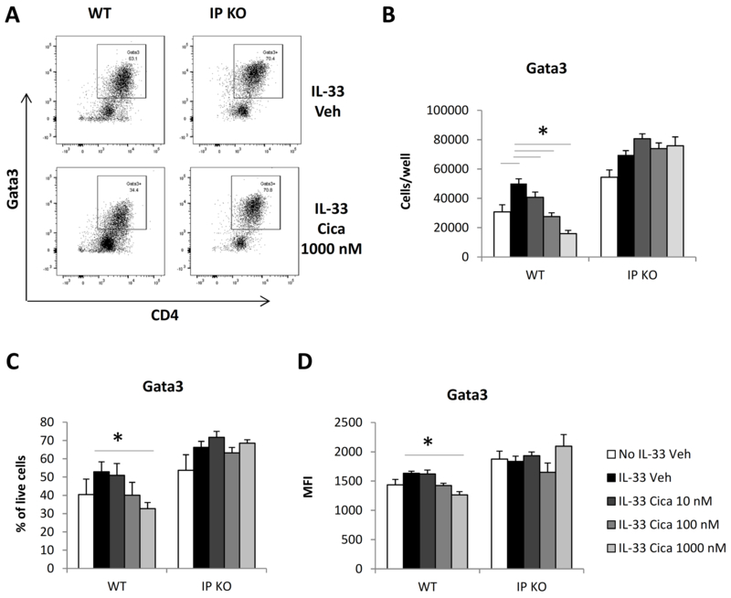
Fig. 3.
Cicaprost decreased Gata3 expression in CD4+ T cells. Naïve CD4+ T cells from WT and IP KO mice were stimulated with anti-CD3 and anti-CD28 Abs in the absence or presence of IL-33 and were treated with vehicle or cicaprost for 3 days. The levels of Gata3 protein expression in the cells was determined by flow cytometry. A. The cells were analyzed for Gata3 expression after gating for lymphocytes and live cells. B. Total numbers of Gata3+CD4+ cells. C. The percentages of Gata3+ CD4+ cells. D. The mean fluorescent intensity (MFI) of Gata3. Data are combined of 3 experiments and presented as mean ± SEM. *, p < 0.05, n=9.

