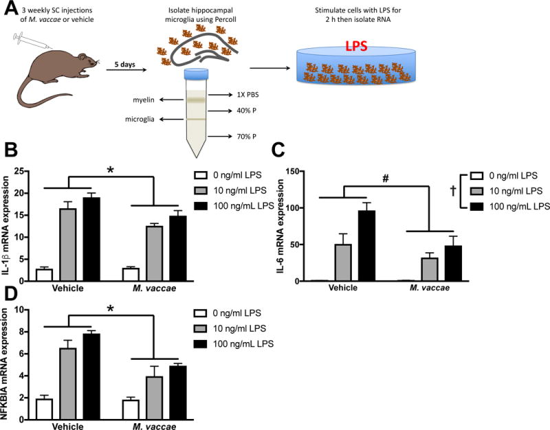
Figure 4. Microglia isolated from aged rats are less inflammatory following in vivo M. vaccae treatment.

(A) Experimental design: aged rats received subcutaneous M. vaccae injections once per week for three weeks. Five days following the final injection, microglia were isolated from the hippocampus and stimulated with LPS. (B) IL-1β, (C) IL-6, and (D) NFKBIA mRNA expression were reduced in microglia isolated from aged M. vaccae-treated rats compared to vehicle-treated rats. Results were analyzed using a 2 × 3 ANOVA with age and LPS as the independent variables (n = 4/group). Data are expressed as mean ± SEM. *LPS × M. vaccae interaction, #main effect of M. vaccae, †main effect of LPS; p < 0.05 in all cases.
