
Figure 1. Type-1 cannabinoid receptors (CB1R) are present on presynaptic terminals, postsynaptic dendrites, and glial cells in the lateral habenula (LHb).
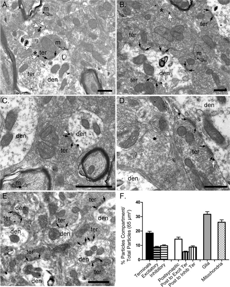
(A-D) Representative electron microscopy images illustrating the presence of CB1R-positive immunogold labeling in the rat LHb. (E) No CB1R labeling was observed in CB1R-KO LHb. Black arrows indicate synapses, small white arrows indicate CB1R-positive immunoparticles on excitatory and inhibitory presynaptic terminals (ter) impinging on postsynaptic dendrites (den), and large white arrows indicate CB1R-positive immunoparticles on glial cells which are indicated with a black asterisk. Scale bars = 500 nm. (F) Bar graph illustrating the percentage of CB1R-positive immunoparticles on excitatory and inhibitory presynaptic terminals (black - horizontal lines), postsynaptic membranes forming synapses with excitatory or inhibitory terminals (white - vertical lines), and glial cells (gray) in the LHb.