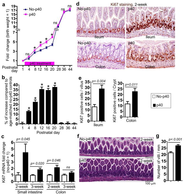
Figure 2. p40 treatment increases growth and epithelial cell proliferation before weaning in wild-type mice.
Mice were treated with p40-containing hydrogels at 0.5, 1, and 1.5 μg/day at postnatal days 2–6, 7–13, and 14–21, respectively. As control, pups were treated with hydrogels without p40 (no-p40). (a) Bodyweight was recorded. The fold change of bodyweight was calculated by comparing the bodyweight at the indicated postnatal day to the bodyweight at birth of the same pup. (b) The percentage of increase = (p40 bodyweight - average of no-p40 bodyweight) / average of no-p40 bodyweight, at the matched postnatal day. (c) Real-time PCR analysis was performed to detect Ki67 gene expression in the small intestine and the colon. The average of mRNA expression levels in 2- and 3-week old mice in the no-p40 group were set as 100%, and the mRNA expression level of each mouse was compared to the average at the same age. (d,e) Ileal and colonic tissues from 2-week old mice were immunostained using an anti-Ki67 antibody and a horseradish peroxidase-conjugated secondary antibody. Slides were developed using DAB and counterstained with hematoxylin. The numbers of positively stained cells are shown. (f–g) Ileal tissues from 2-old mice were prepared for H&E staining and the number of villi per mm is shown. In a–b, no-p40 group: n=15, p40 group: n=17. In c–g: n = 5–7 in each group.

