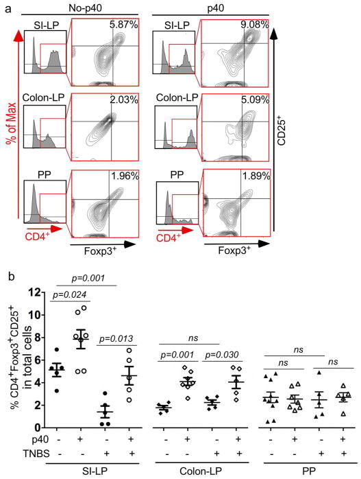
Figure 9. Neonatal p40 treatment promotes induction of intestinal Treg in adult mice.
Wt mice were treated with p40 from postnatal day 2 to day 21, as described in Figure 2. Colitis was induced by TNBS in 7-week old mice, as described in Figure 7. Lymphocytes were isolated from lamina propria (LP) of the small intestine (SI) and the colon and Peyer’s patches (PP). CD4, Foxp3, and CD25 expressing cells were assessed using flow cytometry analysis. Lymphocytes were gated for CD4 and then expression of Foxp3 and CD25 in CD4+ cells were analyzed. (a) Representative CD4 histogram and contour plot of Foxp3 and CD25 are shown. Numbers in Q2 of contour plots represent percentages of CD4+Foxp3+CD25+ in total LP and PP cells. (b) The percentages of CD4+Foxp3+CD25+ cells in total LP and PP cells are shown. In no-p40 SI-LP and colon-LP groups without TNBS, cells from 2 mice/sample. In other groups, cells from 1 mouse/sample.

