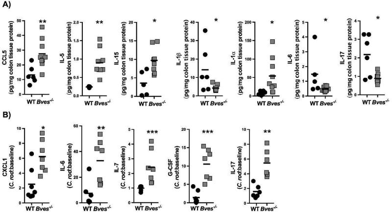
Figure 5. Bves−/− colons display dysregulated chemokine and cytokine expression by Luminex multi-analyte profiling.
(A) Chemokines and cytokines which were significantly different between WT and Bves−/− colon at baseline: CCL5 (**P<0.01, n=7–8), IL-1α (*P<0.05, n=6–8), IL-5 (**P<0.01, n=3–7), IL-1β (*P<0.05), IL-15 (*P<0.05, n=5–6), IL-6 (*P<0.05, n=5–8), and IL-17 (*P<0.05, n=6–8). (B) Chemokines and cytokines which are significantly different between WT and Bves−/− colons after C. rodentium: CXCL1 (*P<0.05, n=7), IL-6 (**P<0.01, n=6–7), IL-7 (***P<0.001, n=6–7), G-CSF (***P<0.001, n=7), and IL-17 (**P<0.01, n=6).

