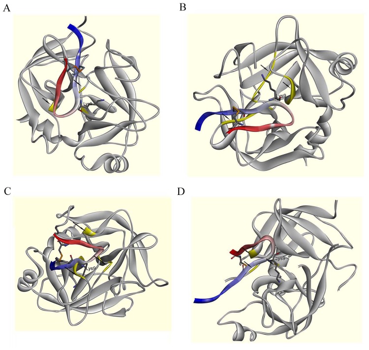
Figure 5.
Structural characteristics of the protease-inhibitor docking output for PE-BBI and KLKs. The secondary structure of PE-BBI was labelled (N-terminal is blue and C-terminal is red), and the active site (Lys9) was shown in stick representation. The disulphide bond was showed in stick representation (Orange). The active site of KLKs were labelled with arrows and highlight (Yellow). (A) Possible interactions model between PE-BBI and KLK4. (B) Possible interactions model between PE-BBI and KLK6. (C) Possible interactions model between PE-BBI and KLK8. (D) Possible interactions model between PE-BBI and KLK10.

