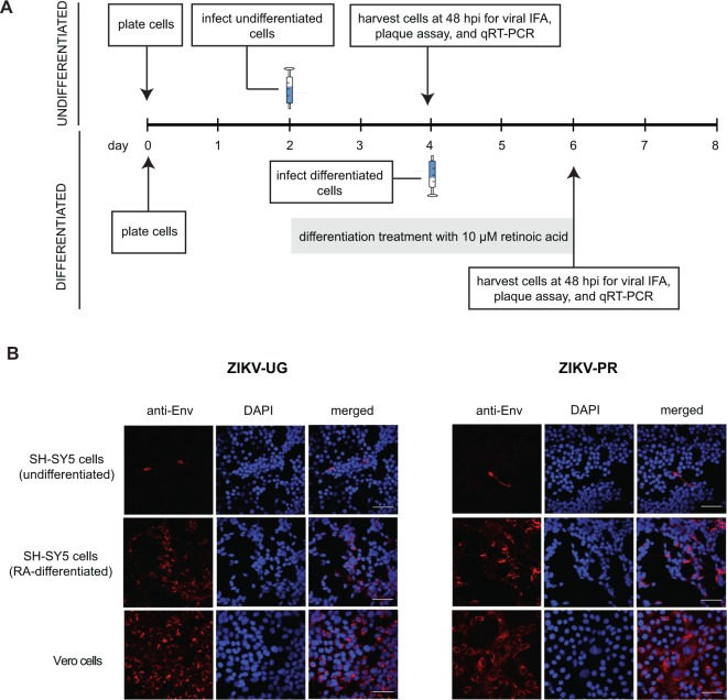
Figure 1.
ZIKV infection of SH-SY5Y neuroblastoma cells. (A) Infection protocol for undifferentiated cells (top) and cells after 2 days of differentiation with retinoic acid (bottom). Syringe icons denote the day of infection with ZIKV. After one hour of incubation, virus was washed twice and collection of time 0 samples was performed. (B) Immunofluorescent staining of SH-SY5Y or Vero cells infected with ZIKV-UG (left) or ZIKV-PR (right) at a multiplicity of infection (MOI) of 1. The 3 columns within each set of 9 panels correspond to staining with a monoclonal antibody against the flaviviral envelope protein (“anti-Env”, staining red, column 1), DAPI stain for cell nuclei (“DAPI”, staining dark blue, column 2), and the merged images (“merged”, column 3). Scale bars represent 50 µm. Abbreviations: IFA, immunofluorescence assay; qRT-PCR, quantitative reverse transcription-polymerase chain reaction; hpi, hours post-infection; DAPI, 4′6-diamidino-2-phenylindole. The images shown are representative images taken from 3 independent experiments, with observation of a minimum of 10 fields under both low (10X) and high (63X) magnification per experiment.

