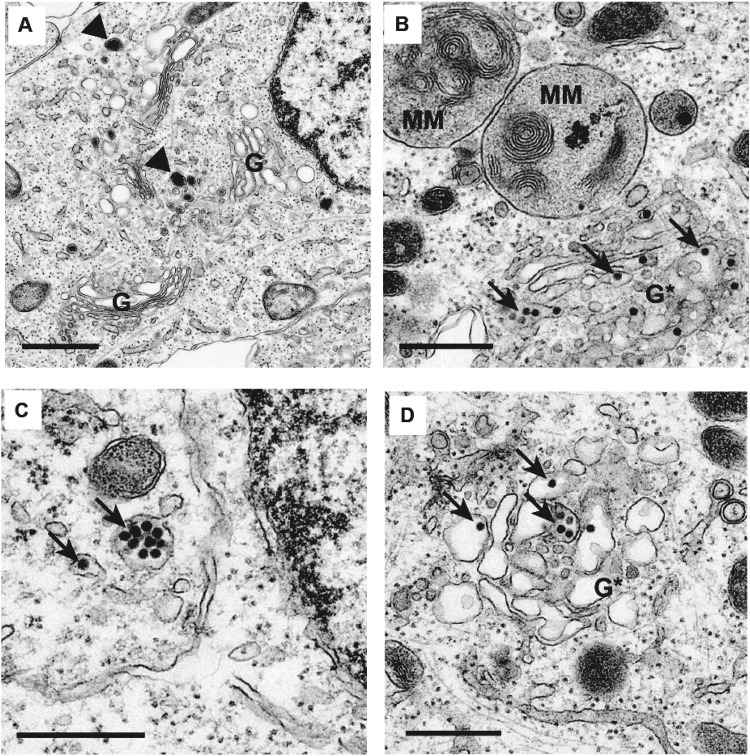
Figure 3.
Transmission electron microscopy of uninfected and ZIKV-infected differentiated SH-SY5Y cells. (A) Uninfected control. Arrowheads (black) show typical neurotransmitter vesicles. (B–D) Differentiated SH-SY5Y cells at 48 hours post-infection with ZIKV-UG. Arrows (black) point to representative viral particles. G, intact Golgi complex; G*, disrupted Golgi complex; MM, multi-membranous areas. Cells are treated with 2 days of retinoic acid prior to ZIKV infection. The scale bar represents 0.5 µm.

